|
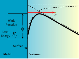
We have pioneered the idea of using functionalized carbon nanotubes (CNT) to enhance thermionic emission, and developed an enhanced thermionic thin film emitter based on carbon nanotubes. This thin film emitter consists of vertically aligned carbon nanotubes grown on a tungsten ribbon surface, and a thin layer of low-work-function emissive materials coating (barium strontium oxide) deposited on top of the carbon nanotubes surface. The barium strontium oxide coating here provides a low work function surface or a low potential barrier for electron emission, and the carbon nanotubes here induce a large field enhancement factor that produces a large Schottky field. The result is an enhanced thermionic emission with significant low effective work function or emission barrier for the electronics. Figures on the right illustrate the structure of the functionalized CNTs and enhanced thermionic emission due to lowering of effective work function. |
|
Growth of Carbon Nanotubes
CNTs were grown on the tungsten ribbon using plasma enhanced chemical vapor deposition (PECVD) technique with the following steps: a) a thin film of nickel, typically about 100 nm, was first sputter deposited on the surface of the tungsten ribbon; b) the film is then etched in NH3 plasma at 600 oC for several minutes; c) this was followed immediately by flowing C2H2 to start the CNT growth; and d) the growth was stopped one the CNTs reach to certain length.
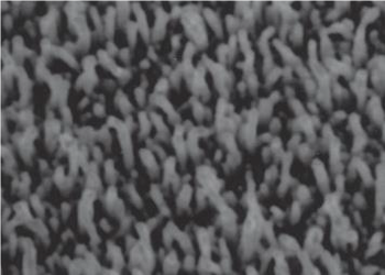
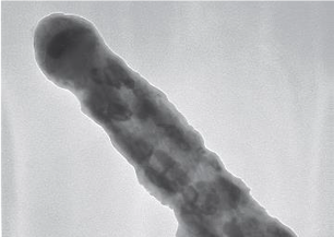
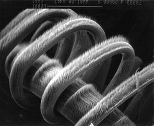
The CNTs were examined with scanning electron microscopy (SEM) and transmission electron microscopy (TEM). Shown on the right are some of the SEM and TEM images of the CNTs grown in the lab.
CNTs were grown on the tungsten ribbon using plasma enhanced chemical vapor deposition (PECVD) technique with the following steps: a) a thin film of nickel, typically about 100 nm, was first sputter deposited on the surface of the tungsten ribbon; b) the film is then etched in NH3 plasma at 600 oC for several minutes; c) this was followed immediately by flowing C2H2 to start the CNT growth; and d) the growth was stopped one the CNTs reach to certain length.
The CNTs were examined with scanning electron microscopy (SEM) and transmission electron microscopy (TEM). Shown on the right are some of the SEM and TEM images of the CNTs grown in the lab.
|
|
(a) and (b) are uncoated CNTs, (c) and (d) are coated nanotubes.
|
